EarSkin and EarCartilage—combining bioengineered human skin with bioprinted cartilage for ear reconstruction
Published by Thamarasee Jeewandara , Medical Xpress, on October 16, 2023
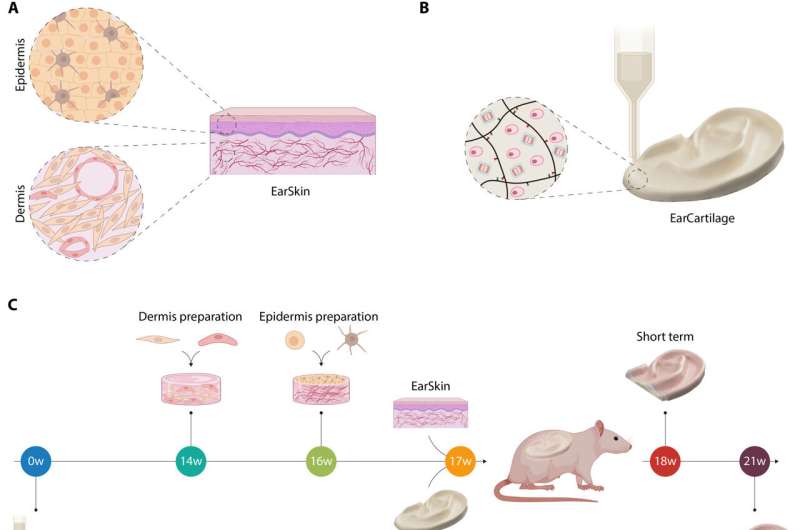
Schematic of the combination of EarCartilage and EarSkin. (A) EarSkin was prepared by creating a dermal layer fabricated out of human fibroblasts and human dermal microvascular endothelial cells (HDMECs) in a collagen I hydrogel. Once matured, an epidermal layer of human keratinocytes and melanocytes was seeded on top of the dermal part. (B) EarCartilage was fabricated using a hyaluronan transglutaminase (HATG)–based bioink together with primary human auricular chondrocytes. Postprinting constructs can be enzymatically crosslinked using calcium-triggered enzymatic crosslinking of factor XIII (30). (C) Experimental timeline of EarCartilage and EarSkin in vivo. EarCartilage was matured for 17 weeks before implantation. EarSkin was created by combining fibroblasts and HDMECs in a collagen I hydrogel and matured for two weeks before seeding keratinocytes and melanocytes on top and culturing it for an additional week. Constructs were then implanted together in vivo and analyzed after one (short term) and four (long term) weeks. w, weeks. (D) Human EarSkin (A) and human EarCartilage (B) were combined in vivo in an immunocompromized rat model. A subcutaneous pocket below the panniculus carnosus was created along the dorsal midline into which EarCartilage was transplanted. The panniculus carnosus was used to cover the EarCartilage framework and to provide rapid nourishment to EarCartilage and EarSkin. EarSkin was then transplanted on top of the panniculus carnosus. Credit: Science Advances, DOI: 10.1126/sciadv.adh1890
Microtia is a congenital disorder that can occur as a malformation of the external ear in children. In a new study published in Science Advances, Dominika Zielinska and a research team in tissue biological research, tissue engineering, polymer technologies and biofabrication at the University of Children's Hospital Zurich, ETH Zurich, Switzerland and the U.S., developed a tissue-engineered treatment approach by using bioprinted autologous auricular cartilage construct as ear cartilage, combined with a bioengineered human pigmented and prevascularized ear skin substrate already tested in immunocompromised rats.
The team confirmed the capacity of the human engineered blood capillaries of EarSkin to connect to the recipient's vasculature within a week for rapid blood perfusion and epidermal maturation. The bioengineered EarSkin demonstrated a stratified epidermis with mature keratinocytes and melanocytes, where the latter resided within the basal layer of the epidermis to efficiently restore the skin color.
Additional in vivo tests showed favorable mechanical stability of the ear cartilage accompanied with improved extracellular matrix deposition. The combined trademarks of Earskin and EarCartilage represented a new treatment approach to microtia, to overcome limits and improve the aesthetic outcomes of microtia reconstruction.


Comments
Post a Comment