Researchers define protocol for high-resolution imaging of living cells using atomic force microscopy
Originally published by Kanazawa Universit at www.phys.org, on September 5, 2023
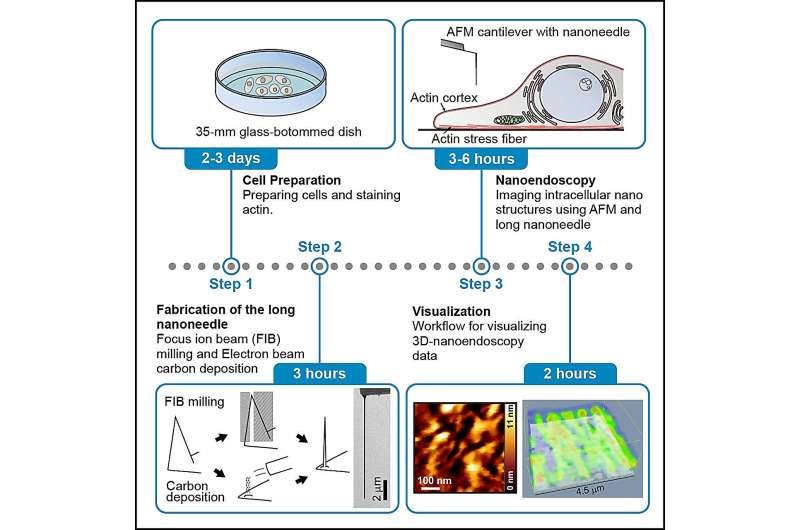
Overview of the method for observing actin fibers in living cells using nanoendoscopy-AFM. Credit: STAR Protocols (2023). DOI: 10.1016/j.xpro.2023.102468
Images of nanoscale structures inside living cells are in increasing demand for the insights into cellular structure and function that they can reveal. So far, the tools for capturing such images have been limited, but researchers led by Takeshi Fukuma and Takehiko Ichikawa at Kanazawa University have now devised and reported a full protocol for using atomic force microscopy (AFM) to image inside living cells. The research is published in the journal STAR Protocols.
AFM was first developed in the 1980s and uses the changes in the forces between a sample surface and a nanoscale tip attached to a cantilever to identify surfaces and produce images of the topography with nanoscale resolution. The technique has grown increasingly sophisticated for extracting information about samples at speeds sufficient for the tool to capture moving images of dynamics at the nanoscale. However, it has so far been limited to surfaces.
Other techniques exist that can provide a view of the inside of a cell, but they have limitations. For instance, there is electron microscopy, which is capable of resolving details at the nanoscale and smaller, but the required operating conditions are not compatible with living cells. Alternatively, fluorescence microscopy is regularly used on living cells, but while fluorescence techniques exist to increase resolution, there are practical challenges that inhibit fluorescence imaging at the nanoscale.


Comments
Post a Comment